The field of mitophagy peptides autophagy research has expanded considerably over the past decade, drawing attention from cell biologists, aging researchers, and performance scientists alike. Autophagy, the cellular process by which damaged or dysfunctional components are broken down and recycled, sits at the intersection of longevity science, metabolic health, and exercise physiology. Mitophagy is a specific, selective branch of autophagy focused entirely on mitochondria. When mitochondria become damaged, fragmented, or inefficient, mitophagy clears them out to make room for healthier ones. Researchers have become increasingly interested in whether peptide compounds can modulate this process, and what that might mean for cellular health.
Understanding this field requires some grounding in why mitochondrial quality control matters. Mitochondria aren't passive energy factories. They're highly dynamic organelles that fuse, divide, and signal to the cell about metabolic status. As cells age or accumulate oxidative stress, mitochondrial dysfunction tends to build up, and the cell's ability to clear dysfunctional mitochondria through mitophagy often declines. Research in model organisms and human tissue samples has pointed toward impaired mitophagy as a common thread in aging-related decline, neurodegenerative conditions, and metabolic dysfunction.
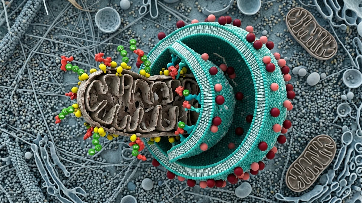
The core machinery of mitophagy depends on a set of proteins that flag damaged mitochondria for degradation. The PINK1/Parkin pathway is one of the most studied. When a mitochondrion loses its membrane potential, PINK1 (a kinase that's normally degraded inside healthy mitochondria) accumulates on the outer membrane. This accumulation recruits Parkin, an E3 ubiquitin ligase, which then tags mitochondrial surface proteins with ubiquitin chains. Autophagy receptors like p62, NDP52, and optineurin recognize those tags and recruit the forming autophagosome, which eventually fuses with a lysosome and digests the contents.
There are also receptor-mediated mitophagy pathways that operate independently of PINK1/Parkin. Proteins like BNIP3L/NIX and FUNDC1 act as direct receptors on the mitochondrial outer membrane, binding to autophagy modifiers and initiating the process without requiring ubiquitin signaling. Each pathway appears to dominate under different physiological conditions, which is one reason this area of research is so complex.
Exercise research touches on this directly. Physical training, particularly endurance exercise, has been shown to upregulate mitophagy pathways in skeletal muscle. This is part of the adaptive response that leads to improved mitochondrial quality and density over time. Some researchers have drawn parallels between exercise-induced mitophagy and the cellular states that certain bioactive peptides appear to influence.
Several peptide compounds have attracted interest for their possible roles in autophagy and mitophagy signaling. These include research peptides that interact with upstream regulators like AMPK and mTORC1, two master switches that tend to act in opposition on autophagy. When AMPK is activated, autophagy tends to be upregulated. When mTORC1 is active, autophagy is typically suppressed. Any peptide that shifts this balance will, in theory, affect mitophagy downstream.
BPC-157 is one compound that has appeared frequently in autophagy-related discussions in the research community. Animal studies have examined its effects on cellular stress responses and tissue recovery, with some researchers speculating that its observed cytoprotective effects may involve autophagic mechanisms. The data remains preliminary and largely limited to rodent models, so drawing broad conclusions would be premature.
Epithalon, a tetrapeptide derived from the pineal peptide preparation Epithalamin, has been studied in the context of aging and cellular longevity. Some researchers have examined whether its reported effects on telomere length and gene expression intersect with mitochondrial quality control pathways. The connection to mitophagy specifically is not yet well-characterized, and researchers working in this space acknowledge that correlation with longevity markers doesn't confirm mechanistic involvement in mitophagy itself.
Humanin, a mitochondria-derived peptide (or MDP), is perhaps the most directly relevant compound in this category. Encoded within the mitochondrial 16S rRNA gene, humanin is secreted by cells and appears to have cytoprotective signaling functions. Research suggests it may interact with IGFBP-3 and BAX pathways to inhibit apoptosis in certain cell types. Some researchers have also examined its relationship to mitochondrial biogenesis and quality surveillance, though isolating its specific role in mitophagy versus broader mitochondrial protection is an active area of investigation.
The relationship between AMPK activation, mTOR suppression, and autophagy induction is one of the most studied regulatory mechanisms in cellular biology. Caloric restriction, exercise, and certain pharmacological compounds like rapamycin and metformin all converge on this axis. Researchers studying peptides for autophagy modulation are asking whether specific short-chain amino acid sequences can mimic or amplify some of these effects with more tissue-specific precision.
The theoretical appeal is straightforward. Small molecules that activate AMPK broadly may create metabolic side effects across multiple tissue types. A peptide that interacts with a more upstream or localized signal could, in theory, influence autophagy in a targeted way. Whether current research peptides actually achieve this specificity in vivo is not yet clear from the available data.
One limitation worth naming directly: most peptide research showing autophagy effects has been conducted in vitro or in rodent models. The translation from a cell culture result to a meaningful effect in a living human is not automatic, and many compounds that look promising in early models don't replicate the same results in clinical settings. This doesn't mean the research is without value, but it does mean that enthusiasm should be calibrated to the actual stage of evidence.
One reason mitophagy peptides autophagy research has gained traction outside purely academic circles is the link between mitochondrial quality and physical performance. Athletes and coaches interested in recovery science have noted that mitochondrial turnover plays a role in how well muscle tissue adapts to training stress. Impaired mitophagy in aging muscle, a phenomenon some researchers call mitochondrial crowding, has been proposed as a partial explanation for the decline in aerobic capacity observed with age.
The connection to recovery peptide research is natural here. Compounds studied for their role in tissue repair, like growth hormone-related secretagogues and IGF-1 pathway modulators, overlap with systems that also regulate mitochondrial dynamics. Related subjects like NAD+ precursor research and sirtuins (SIRT1 and SIRT3 are particularly tied to mitophagy regulation) have also entered this conversation, since sirtuin activity is known to influence both mitophagy signaling and mitochondrial biogenesis.
Researchers studying GLP-1 pathway peptides have separately noted mitochondrial effects in metabolic tissues, particularly in the context of insulin sensitivity and pancreatic beta cell function. Whether these mitochondrial effects involve mitophagy specifically, or represent more general mitochondrial protection, is an open question that current research hasn't fully resolved.
Evaluating claims in this field requires some understanding of the tools researchers use to measure mitophagy. Fluorescent reporter systems like mt-Keima and mito-QC allow researchers to track mitochondria as they enter lysosomes. Western blotting for LC3-II lipidation and p62 degradation provides indirect evidence of autophagic flux. Transmission electron microscopy can visualize autophagosomes containing mitochondrial content. These methods each have limitations, and results can vary depending on the model system used.
When reviewing peptide studies that claim to show mitophagy effects, it's worth checking which of these methods were actually used. A study showing reduced oxidative stress markers or improved mitochondrial membrane potential is not the same as a study demonstrating increased autophagic flux with mitochondrial cargo. The distinction matters because autophagy induction and mitochondrial protection can happen through entirely separate mechanisms.
According to practitioners and researchers active in this space, the most scientifically credible signals currently exist for compounds that interact with known autophagy nodes like AMPK, ULK1, or the Beclin-1 complex. Peptides with documented interactions at these sites have at least a mechanistic framework to support autophagy-related hypotheses. Compounds where the proposed mechanism is vague or indirect require much more caution before any conclusions are drawn.
The honest state of the field is one of genuine scientific interest combined with significant gaps in human data. That's not a failure of the research, it's an accurate description of where things stand. Early-stage science often looks messier than popular accounts suggest, and mitophagy peptide research is no exception.
For those following this field from a health optimization or performance science perspective, the most useful frame is probably one of informed patience. The biological rationale for targeting mitophagy is sound. Mitochondrial quality control is genuinely central to cellular aging, metabolic function, and physical capacity. The question of whether peptide compounds can meaningfully modulate this process in humans, safely and reliably, remains open.
Lifestyle factors with established mitophagy-inducing effects include endurance exercise, intermittent fasting, and heat stress via sauna protocols. These don't carry the same regulatory and safety uncertainties as experimental peptides, and they have substantially more human data behind them. Researchers studying peptides in this space would likely agree that these foundational inputs remain the most well-supported levers available to non-clinical populations.
Interest in compounds like humanin, BPC-157 variants, or novel mitochondria-targeting peptides is scientifically legitimate. Treating preliminary animal data as confirmed human application is not. The gap between those two positions is where careful reading of the literature becomes essential.
This article is for informational and research purposes only and does not constitute medical advice. The compounds discussed are experimental and have not been approved by the FDA or equivalent regulatory bodies for human therapeutic use. Nothing in this article should be interpreted as a recommendation to use, obtain, or administer any compound described. Always consult a qualified healthcare professional before making any changes to your health practices. For research purposes only — not medical advice.