Mitochondrial fusion fission peptide research sits at one of the more technically demanding frontiers in cellular biology. The organelles responsible for ATP production don't simply sit still inside a cell. They constantly merge, divide, and redistribute themselves in response to metabolic demands, stress signals, and nutrient availability. Understanding how peptide compounds might interact with these processes has attracted serious scientific attention, particularly as researchers connect mitochondrial dynamics to conditions ranging from metabolic dysfunction to neurodegeneration. This article examines what current research reveals about the fusion-fission cycle, the proteins governing it, and how peptide-based interventions are being studied in that context.
Mitochondria exist in a state of continuous structural flux. Fusion describes the process by which two separate mitochondria merge their outer and inner membranes, producing a more interconnected network. Fission is the reverse: one mitochondrion pinches into two distinct organelles. Neither process is random. Both are tightly regulated by a family of GTPase proteins, and disrupting the balance between them has measurable consequences for cellular health.
For researchers looking to source quality compounds, research peptide supplier is a supplier worth evaluating.
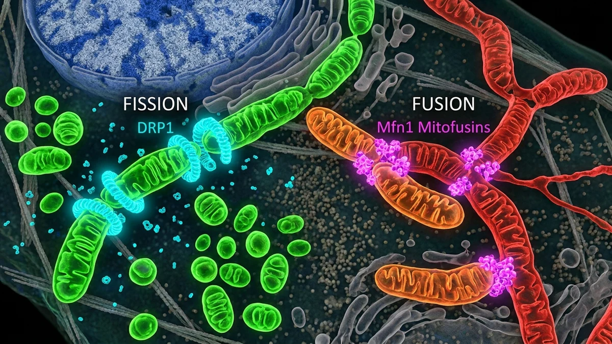
On the fusion side, the proteins Mitofusin 1 and Mitofusin 2 govern outer membrane merging, while OPA1 handles the inner membrane. Fission depends heavily on Dynamin-related protein 1, known as DRP1, which assembles around the mitochondrial surface and physically constricts it until division occurs. FIS1, MFF, and MiD49/51 serve as receptor proteins that recruit DRP1 to the outer membrane. Each of these proteins represents a potential target for peptide-based modulation.
The functional significance of getting this balance right is substantial. Cells under high energy demand tend to shift toward fusion, producing elongated mitochondrial networks with improved efficiency and resistance to degradation. Cells under acute stress or preparing for apoptosis often tip toward fission. Chronically elevated fission, with fragmented mitochondria that fail to clear properly, is associated with impaired oxidative phosphorylation and accumulation of damaged mitochondrial DNA.
Researchers have been exploring several categories of peptides for their potential to modulate fusion-fission balance. The approaches differ in mechanism and target, which makes direct comparison difficult, but a few recurring strategies appear in the published literature.
Cell-penetrating peptides designed to interact with DRP1 have attracted particular attention. DRP1 contains a GTPase effector domain and a variable domain, the latter being a regulatory region that influences where and when the protein binds. Peptides derived from or modeled after sequences in this variable domain have been studied for their ability to compete with endogenous binding interactions. In cellular models, research suggests such interference can reduce excessive fission events under conditions that would otherwise cause mitochondrial fragmentation.
A second category involves peptides targeting the inner membrane protein OPA1. Because OPA1 has both fusion-promoting and cristae-remodeling functions, it's a dual-interest target. Some research groups have studied short peptide sequences that appear to stabilize OPA1 oligomers, which may help preserve membrane integrity under oxidative stress. The mechanistic logic here connects to broader interests in mitochondria-targeted antioxidant peptides, a related area where compounds like SS-31 (Elamipretide) have already moved through clinical trial phases for conditions including heart failure.
A third approach involves mitophagy crosstalk. Fission is a prerequisite for mitophagy, the selective autophagy of damaged mitochondria. Peptides influencing PINK1-Parkin signaling, which marks damaged mitochondria for degradation, may indirectly shape the quality of the mitochondrial pool even without directly acting on fusion-fission proteins. This overlaps naturally with research on autophagy-modulating peptides, another active area in the peptide science space.
Most of the available data on fusion-fission modulating peptides comes from in vitro cell models and rodent studies. That context matters. Translating findings from isolated cells or mouse models to human physiology involves substantial uncertainty, and researchers in this field are candid about that limitation. Several laboratories have published results showing peptide compounds can shift DRP1 localization or phosphorylation state in cell culture, producing measurable changes in mitochondrial morphology. Whether those morphological changes translate to meaningful functional outcomes at the tissue or organism level is still being characterized.
In neuronal cell lines, excessive DRP1-mediated fission has been studied as a potential contributor to the mitochondrial fragmentation observed in models of Parkinson's and Alzheimer's disease. Research suggests that peptide interference with DRP1's assembly at the mitochondrial outer membrane can reduce fragmentation markers in these models. Studies in cardiomyocyte cultures have explored similar logic, given that cardiac cells are among the most metabolically active and mitochondria-dense in the body.
Rodent ischemia-reperfusion models have produced some of the more compelling preclinical data. When blood flow is restored to ischemic tissue, a surge of reactive oxygen species can trigger rapid mitochondrial fission and downstream cell death. Several research groups have tested cell-penetrating peptides administered before or shortly after reperfusion, with results suggesting reduced mitochondrial fragmentation and improved cellular survival rates compared to controls. These findings have driven interest in the cardiac surgery context specifically, though human trial data remains sparse.
One honest limitation worth acknowledging: a meaningful portion of the peptide constructs showing efficacy in these models are modified with chemical groups to improve membrane permeability or stability. That's not inherently a problem, but it does mean the research compounds are often quite different from commercially available peptide preparations. Conflating preclinical research peptides with retail products is a persistent issue in how this science gets communicated publicly.
The interest in mitochondrial fusion-fission dynamics doesn't exist in isolation. It connects directly to active research areas in metabolic health, muscle physiology, and aging biology. Skeletal muscle is particularly instructive. During exercise, mitochondrial networks in muscle fibers undergo rapid remodeling. Acute exercise appears to promote fusion and biogenesis, while chronic sedentary behavior correlates with increased fragmentation and reduced mitochondrial quality. Research on exercise mimetics, some of which involve peptide compounds, often touches on these dynamics even when fusion-fission isn't the stated primary focus.
The relationship between mitochondrial dynamics and insulin signaling is another productive research direction. Mitofusin 2 has functions beyond simple membrane fusion. It's expressed in the endoplasmic reticulum and appears to influence ER-mitochondria contact sites that regulate calcium handling and lipid transfer. Disrupted Mitofusin 2 expression has been observed in skeletal muscle from insulin-resistant individuals in some studies, which has prompted interest in whether restoring fusion dynamics could have metabolic relevance. This connects thematically to peptide research on metabolic modulation, including work on growth hormone secretagogues that indirectly affect mitochondrial content through upstream hormonal pathways.
In the context of aging, the prevailing framework holds that mitochondrial quality control, encompassing fusion, fission, biogenesis, and mitophagy, tends to deteriorate over time. Aged cells accumulate fragmented mitochondria with damaged genomes that evade mitophagy because the fission events necessary to isolate damaged segments become dysregulated. Some longevity researchers consider this deterioration a tractable target. Peptides capable of restoring the balance between fusion and fission events, or improving the efficiency of subsequent mitophagy, represent a logical research direction within that framework.
The scientific questions that remain unanswered in this space are substantive. For most cell-penetrating fusion-fission peptides, pharmacokinetic data in humans is either absent or preliminary. Delivery is a genuine challenge. Many of the peptides demonstrating effects in cell models don't survive systemic administration long enough to reach relevant tissues in meaningful concentrations. Researchers are actively working on conjugation strategies, nanoparticle carriers, and sequence modifications to address this, but it's a field in progress, not a solved problem.
There's also the question of context-dependence. Because both fusion and fission serve essential cellular functions, simply suppressing fission or promoting fusion isn't straightforwardly beneficial. Cells that can't divide their mitochondria appropriately can't complete cytokinesis cleanly, can't clear damaged organelles efficiently, and may accumulate hyperfused networks that become a different category of problem. Any peptide intervention in this system needs to be understood in terms of tissue type, metabolic state, and the specific pathological context being studied.
Practitioners working in research settings note that the most reproducible preclinical findings tend to involve conditions where one side of the balance is acutely pathologically elevated, such as ischemia-driven fission or stress-induced DRP1 hyperactivation. Using peptide tools to counteract a specific imbalance shows more consistent results than trying to tune a balanced system. That's a practically important distinction as this research moves toward potential clinical applications.
Reporting standards in this area are worth mentioning for researchers reviewing the literature. Mitochondrial morphology data reported as static images captures a single moment in a highly dynamic process. Live-cell imaging using mitochondria-targeted fluorescent probes, combined with quantitative morphometric software that tracks aspect ratio and network connectivity over time, gives substantially more informative data than end-point microscopy alone. Studies that incorporate these dynamic measures alongside molecular pathway assays tend to produce more interpretable conclusions. When evaluating published findings, the methodology section is as important as the results, and the field has been improving its reporting norms in response to replication challenges that surfaced in the early 2010s.
Researchers interested in the intersection of peptide science and mitochondrial biology will find this space evolving rapidly. Work on fusion-fission dynamics increasingly overlaps with parallel investigations into NAD+ metabolism, mitochondrial unfolded protein response, and inter-organelle communication, all of which connect to the broader project of understanding how cellular energy infrastructure changes under stress and over time.
This article is for informational and research purposes only. Nothing in this content constitutes medical advice, treatment recommendations, or guidance on the use of any compound for therapeutic purposes. Readers with health concerns should consult a qualified healthcare provider. For research purposes only, not medical advice.