Cardiolipin targeting peptides mitochondrial research has become one of the more specific and technically demanding frontiers in cellular biology. Cardiolipin itself is a phospholipid found almost exclusively in the inner mitochondrial membrane, and its structural integrity appears closely tied to how well mitochondria generate energy, manage oxidative stress, and initiate apoptotic signaling. When cardiolipin is oxidized or its distribution within the membrane becomes disrupted, downstream effects ripple across multiple cellular processes. Peptides designed to interact with cardiolipin represent an attempt to study and potentially modulate these processes at a precise, membrane-level scale.
The interest in this area isn't accidental. Mitochondrial dysfunction sits at the intersection of aging research, metabolic health, neurodegenerative disease models, and exercise physiology. Cardiolipin's role as a structural scaffold for the electron transport chain complexes makes it a logical focal point. When researchers look at how energy production falters under conditions of chronic inflammation or cellular aging, cardiolipin oxidation consistently appears in the picture.
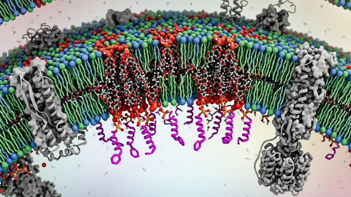
Cardiolipin's molecular structure is unusual. Unlike most phospholipids, it carries four fatty acid tails and two phosphate groups, which gives it a conical shape that promotes the high curvature of the inner mitochondrial membrane. That curvature isn't cosmetic. It's thought to be essential for efficient electron transport chain assembly and for the formation of cristae, the folded inner membrane structures where ATP synthesis occurs.
Research suggests that cardiolipin directly stabilizes complexes I, III, IV, and the ATP synthase within the electron transport chain. It acts almost like a molecular glue, holding these large protein complexes in position and facilitating their organization into supercomplexes called respirasomes. When cardiolipin content drops or its fatty acid composition shifts due to oxidative damage, supercomplex stability appears to decline, which correlates with reduced respiratory efficiency.
There's also a signaling dimension. Under conditions of cellular stress, cardiolipin migrates from the inner membrane to the outer mitochondrial membrane, where it becomes a recognition signal for the autophagy machinery. This process, specifically mitophagy, allows damaged mitochondria to be cleared. Cardiolipin's translocation appears to serve as a kind of quality-control flag, and peptides that interact with it have been used in research settings to probe exactly how this signaling sequence unfolds.
One acknowledged limitation across this field is that most cardiolipin research has been conducted in cell culture or animal models. Translating these findings to human physiological contexts remains an active and sometimes contentious process.
Peptides designed for cardiolipin interaction typically exploit electrostatic and hydrophobic properties. Cardiolipin carries a net negative charge at physiological pH due to its phosphate groups, which makes it a natural binding partner for cationic, or positively charged, peptides. Many of the peptides studied in this space are small, often four to ten amino acids in length, and are designed to localize specifically to mitochondria rather than distributing broadly across cellular membranes.
One of the more studied frameworks involves what researchers call Szeto-Schiller (SS) peptides, a class of tetrapeptides developed to accumulate preferentially in the inner mitochondrial membrane. Their alternating aromatic and cationic amino acid pattern appears to drive both mitochondrial targeting and cardiolipin binding. In research settings, these peptides have been used to study how protecting cardiolipin from oxidative modification affects respiratory function and cell survival under stress conditions.
The design logic here is worth unpacking. Mitochondria maintain a strongly negative membrane potential across the inner membrane, somewhere in the range of negative 150 to 180 millivolts, which creates an electrochemical gradient that pulls cationic molecules inward. Peptides engineered with the right charge distribution can exploit this gradient to achieve concentrations inside mitochondria that far exceed their concentrations in the surrounding cytoplasm. This selective accumulation is what makes them useful as research tools.
It's also why researchers have become interested in using these peptides to study topics like mitochondrial reactive oxygen species production, which connects directly to research on oxidative stress and cellular aging. The peptides can be used as probes or as pharmacological tools in controlled experimental conditions to dissect cause-and-effect relationships that are otherwise difficult to isolate.
Mitochondrial oxidative stress doesn't develop in isolation. It's woven into broader conversations about aging biology, metabolic function, and longevity research. Cardiolipin is particularly vulnerable to oxidation because it's located so close to where reactive oxygen species are generated during electron transport. Its polyunsaturated fatty acid tails, especially when they're enriched in linoleic acid, are susceptible to lipid peroxidation.
Research in aged animal models has documented consistent declines in cardiolipin content and changes in its fatty acid profile compared to younger counterparts. Some researchers argue that this isn't simply a consequence of aging but may be a contributing mechanism. The logic is straightforward: if cardiolipin oxidation disrupts supercomplex stability, and supercomplex disorganization reduces respiratory efficiency, then a cycle can develop where declining mitochondrial output generates more oxidative stress, which damages more cardiolipin.
Cardiolipin targeting peptides have been used in research settings to test whether interrupting this cycle at the cardiolipin level changes outcomes in aged cell models and in certain animal models of age-related decline. This connects to broader research areas, including work on NAD+ metabolism and mitochondrial biogenesis, since all of these pathways ultimately feed into the same cellular energy economy.
There's no shortage of open questions. The field is still working out which specific cardiolipin species are most functionally relevant, how tissue-specific cardiolipin composition affects how cells respond to these peptides, and whether the protective effects seen in model systems translate meaningfully to more complex biological contexts.
The brain's relationship with mitochondrial function is especially intimate. Neurons are post-mitotic, highly energy-dependent cells that can't simply divide to replace damaged counterparts. Mitochondrial health in neurons has therefore attracted serious attention in the context of neurodegenerative disease research.
Cardiolipin abnormalities have been documented in research models of Parkinson's disease, Alzheimer's disease, and traumatic brain injury. In Parkinson's research specifically, there's substantial interest in how the protein alpha-synuclein interacts with cardiolipin, since some research suggests that pathological forms of alpha-synuclein can bind to cardiolipin and disrupt its distribution within the mitochondrial membrane.
Cardiolipin targeting peptides have been used in this context as investigative tools, helping researchers understand whether preserving cardiolipin integrity affects mitochondrial morphology, neuronal survival under oxidative challenge, or downstream inflammatory signaling in microglia and astrocytes. This line of inquiry connects directly to the broader area of mitochondria-targeted peptide research, which has expanded considerably over the past decade as better tools for visualizing and quantifying mitochondrial membrane dynamics have become available.
The work is painstaking and the translation challenges are real. Brain tissue has its own lipid composition, its own antioxidant environment, and its own metabolic demands. Findings from one model system don't automatically port over to another, which is why researchers in this space tend to be careful about generalizing too quickly from cellular or rodent data.
Mitochondrial adaptation to exercise is one of the most well-characterized phenomena in sports science. Endurance training consistently increases mitochondrial density, improves electron transport chain efficiency, and alters mitochondrial membrane composition. What's less commonly discussed is how cardiolipin composition changes in response to physical training.
Research suggests that exercise-induced mitochondrial biogenesis in skeletal muscle is accompanied by changes in cardiolipin content and, in some studies, in cardiolipin's fatty acid profile. Whether this is a driver of improved respiratory function or a downstream consequence of mitochondrial proliferation isn't fully resolved. Some researchers have hypothesized that the cardiolipin remodeling that occurs with training represents a form of membrane-level optimization, aligning mitochondrial structure more tightly with the energy demands that regular exercise imposes.
Cardiolipin targeting peptides have entered exercise physiology research largely as mechanistic tools, used to probe how cardiolipin oxidation during high-intensity exercise affects recovery, mitochondrial quality control, and the signaling cascades that drive adaptation. This intersects with research on cellular stress responses and the role of mitophagy in removing exercise-damaged mitochondria so they can be replaced with higher-quality ones.
From a practical standpoint, this research doesn't yet translate into specific protocols or recommendations. What it does offer is a more granular understanding of why mitochondrial health responds so well to consistent aerobic training, and what the membrane-level mechanisms underlying that response actually look like.
The field is moving toward greater specificity. Early cardiolipin targeting peptide research focused on whether these molecules could accumulate in mitochondria and whether that accumulation correlated with measurable changes in function. Current work is more nuanced, asking which specific cardiolipin species are most critical under which conditions, how mitochondrial network dynamics (fusion, fission, and mitophagy) respond to cardiolipin-level interventions, and whether tissue-specific formulation strategies might make these tools more useful across a wider range of research applications.
There's also growing interest in combining cardiolipin-targeted approaches with other mitochondria-focused research areas, including work on peptides involved in mitochondrial biogenesis signaling and compounds that interact with the electron transport chain more broadly. The idea is to build a more complete mechanistic picture rather than studying individual pathways in isolation.
What the existing body of research makes reasonably clear is that cardiolipin is not a passive structural element in the mitochondrial membrane. It's an active participant in energy metabolism, stress signaling, and cell fate decisions. Peptides designed to interact with it have proven to be valuable instruments for studying those roles, even if significant questions about mechanism and translation remain open.
This article is for informational and research purposes only. Nothing in this article constitutes medical advice, diagnosis, or treatment recommendations. The peptides and research topics discussed are intended for scientific and educational reference. Consult a qualified healthcare professional before making any decisions related to health, supplementation, or therapeutic interventions. For research purposes only — not medical advice.